Skin Layers Explained: Epidermis, Dermis and Hypodermis
Skin layers explain where moisturizer goes and what each part does. See how the epidermis, dermis and hypodermis shape your daily product order.

Think about the moment you press a new moisturizer into your cheeks and wonder where it actually goes. Skin isn’t one flat sheet. It’s a stack of layers, each with its own job, and knowing them makes your routine easier to build.
Here’s the short answer: skin has three main layers, the epidermis, the dermis and the hypodermis, and each does different work.
In this post, we’ll walk through them, the special cells that live in them, and what changes as skin ages, then show how that shapes what you put on first, second and last.
Skin Layers and What Your Skin Does
Your skin is part of the integumentary system, the body’s outer covering system that also includes hair and nails. Before we start layering products, here’s what your skin is already doing for you.
Why skin is the body’s largest organ
According to Anatomy, Skin (Integument), skin is the body’s largest and primary protective organ. It covers the entire exterior and serves as a first physical barrier against the environment.
SEER Training’s Anatomy of the Skin says it’s about 2 mm thick on average and weighs roughly six pounds, which is a lot of real estate for one organ.
How skin protects you every day
Skin doesn’t just cover you. It works for you all day, and its jobs include:
- Acting as a barrier against pathogens, the germs that can make you sick
- Shielding you from heat, light, and injury
- Regulating body temperature
- Gathering sensory information so you can feel what you touch
- Storing water, fat, and vitamin D
- Playing a role in your immune system
Think of a bridesmaid standing outside for an hour of group photos.
Her skin takes the heat and light the whole time, so a few minutes of care before she leaves the house helps her stay comfortable and still look like herself in the pictures. Your skin is always on duty.
How thin and thick skin differ
Skin isn’t the same everywhere. Thin, hairy skin is the most common type, while thick, hairless skin shows up in friction areas like the palms and soles. Color and texture vary too, so your face, hands, and feet may look and feel different.
That’s your first clue for layering: one product rarely suits every area equally.
Different Ways to Count Skin Layers
Skin doesn’t come with a single official count. Slice it coarsely and you get three layers; slice it finely and you get seven. Here’s how both work.
The simple three-layer breakdown
The simplest model splits skin into three layers:
-
the outer epidermis
-
the middle dermis
-
the deepest hypodermis
-
Epidermis: the thin outer layer you see and touch, which keeps moisture in and irritants out.
-
Dermis: the thicker middle layer that gives skin its structure, along with blood vessels and nerves.
-
Hypodermis: the deepest layer, mostly fat, which cushions and insulates.
The layers differ in function and thickness. The epidermis is the thinnest, but it does a lot of the protecting.
For example, a bride doing her own makeup at 6 a.m. works almost entirely with the epidermis, so a light, comfortable base there keeps her foundation smooth through the ceremony.
Finer breakdowns into five or seven layers
Finer models split the same skin into more pieces. The epidermis alone is often divided into five strata, or sublayers. Listed top to bottom:
- Stratum corneum: the flat, dead cells at the surface, where moisturizer and sunscreen sit.
- Stratum lucidum: a thin, clear band found mainly on palms and soles.
- Stratum granulosum: where cells flatten and begin to harden toward the surface.
- Stratum spinosum: cells with spiny-looking edges that hold together and keep the epidermis sturdy.
- Stratum basale: the deepest epidermal layer, where new skin cells form.
Add the two dermal layers, the upper papillary layer and the deeper reticular layer, and you land on seven. Same skin, different count. Most skincare happens at the top, but the layers beneath do the building.
What Is the Toughest Layer of the Epidermis?
The toughest layer of the epidermis (the thin outer skin you live in) is the stratum corneum. It’s tough because of what it does, not how thick it is, and it’s more fragile than that toughness suggests.
Here’s how it’s built, what sits beneath it, and why it needs a gentle hand.
How the top layer is built and sealed
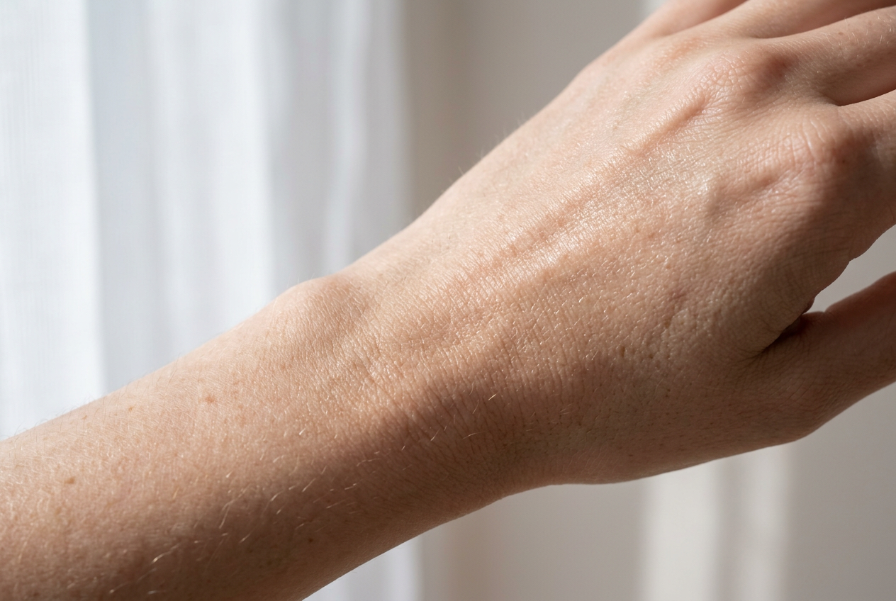
The stratum corneum is the outermost visible layer, the part of your skin you see and touch every day. Its cells are squamous, meaning flat and scale-like, and they fit together into a waterproof barrier.
That same seal blocks germs and infection from getting in. Thickness varies by location, so the top layer on one part of your body isn’t identical to the top layer on another.
How melanin sets your skin tone
Melanocytes are the cells that produce melanin, the pigment that determines your complexion. Melanin also gives your skin some natural sun protection, which makes it a useful ally.
A study on how ultraviolet (UV) light drives pigmentation found that UV light switches up the gene for tyrosinase, the enzyme that drives melanin production.
That’s why melanocytes ramp up with sun exposure, and it’s a good reason to make sun protection a step you never skip.
How skin cells renew and shed
Your skin is always replacing itself. A study on how epidermal cells renew themselves describes the epidermis as a major site of self-renewal. New cells are made in the basal layer, migrate upward toward the surface, and eventually slough off.
Researchers can measure both the turnover time and the shedding amount of that cycle.
Layers inside the epidermis, top to bottom
From the surface down, the epidermis has five layers:
- Stratum corneum: the surface layer we just covered.
- Stratum lucidum: the layer directly beneath it.
- Stratum granulosum: the layer below that.
- Stratum spinosum: the next one down.
- Stratum basale: the bottom layer, where new cells are made.
Why the stratum corneum is so fragile
Tough doesn’t mean indestructible. The stratum corneum is made of dead, flattened keratinized cells (cells that have hardened into a protective shell), and it’s a very thin outer layer with little to spare. Harsh exfoliators can damage it.
Once it’s damaged, moisture escapes, leaving skin dry and tight.
Consider a bride who scrubs her face with a harsh exfoliator the week before her wedding. By the ceremony her skin feels tight and dry, and her makeup looks patchy in the photos. A gentler routine from the start would have spared her that scramble.
That’s why light layers matter. A thin, well-chosen layer can help protect the barrier without piling on more than your skin needs.
Topical products (anything you apply to the skin) reach this layer first, so it’s the starting point for how far they can penetrate.
Where Is the Dermis Located in the Skin?
The dermis sits directly beneath the epidermis, the outer layer you see and touch every day. It does much of its work out of sight, so let’s look at what’s inside and what it does.
What lives inside the dermis
Several structures are packed into the dermis:
- Nerve endings
- Sweat and oil glands
- Hair follicles
- Blood vessels
How fibroblasts build collagen and elastin
Fibroblasts are the cells that produce the dermis’s fibers. They make collagen, the fiber that gives skin its strength, and elastin, the fiber that gives it elasticity. The dermis also holds hyaluronic acid, a substance that holds water.
So what causes skin aging? One part of the answer is collagen loss.
According to Eklouh-Molinier et al., with aging, both the quality and quantity of type I collagen in the dermis decrease, which leads to more wrinkles, drier skin, and lower moisture capacity. Less collagen, more wrinkles.
UV damage reaches the dermis too, not just the surface.
Upper and lower zones of the dermis
The dermis has two zones:
- Papillary layer: the thin upper zone, where fingerprints originate.
- Reticular layer: the thick lower zone, full of dense collagen bundles.
Your fingerprints come from the upper zone, not the lower one.
What the dermis does for your body
The dermis does a lot of jobs. Its sweat glands produce sweat, which helps with cooling. Nerve endings handle touch, temperature, and pain sensation.
The dermis also supplies blood to the epidermis, and capillaries, the smallest blood vessels, help with temperature regulation. When skin gets hurt, the dermis works as a regeneration reservoir.
Consider a bridesmaid who nicks her thumb on a pin during the final fitting. Bleeding occurs when skin is cut, and that’s the dermis’s blood supply at work. It’s a small reminder that the dermis keeps working, even when you can’t see it.

Deep Fat Layer Known as the Hypodermis
Here’s the layer you’ll never see in the mirror, but it’s busy behind the scenes.
What the deepest layer is made of
The hypodermis is the deepest layer of the skin, and it’s also called the subcutis. It’s built mostly from fat and connective tissue, the fibrous tissue that holds the body’s structures together. Think of it as the padding everything above it rests on.
How this layer cushions, insulates and anchors
This layer is less about surface and more about support. It does a few key jobs:
- Anchoring. It attaches skin to the muscle and bone beneath it.
- Padding. It helps protect deeper tissue against injury from bumps and pressure.
- Insulation and temperature regulation. Its fat helps keep the body warm and the temperature steady.
- Pathways. Nerves and blood vessels pass through this layer on their way to the skin above.
Consider a bride standing through a long, breezy outdoor ceremony. The insulating fat under her skin helps hold in warmth, so the chill is less likely to pull her focus from her vows. That’s a lot of quiet work.
Special Skin Cells and What They Do
Some specialized cells and structures do jobs beyond holding skin together.
Cells that alert your immune system
Langerhans cells live in the epidermis, the skin’s outer layer, and act as sentinels for your immune system. Dendritic cells in the stratum spinosum, a middle layer of the epidermis, share this role.
Part of the job is surveying for abnormalities in deoxyribonucleic acid (DNA), the genetic instructions inside each cell, which can signal trouble. It’s about catching problems before they show.
Receptors that sense touch and pressure
Several receptors and nerves pick up touch and pressure:
- Merkel cells sit near the surface and respond to light, steady touch.
- Pacinian corpuscles sit deeper and respond to pressure and vibration.
- Basket cells wrap around each hair and register when it moves.
- Sensory nerves carry these signals to the brain, so you can tell a tap from a squeeze.
Consider a bride fastening her veil at the mirror. The light touch at her temples and the pressure of each comb reach her brain through these receptors, so she can place every pin without looking. Bring that same lightness to the sink.
Glands, hair and what makes goosebumps
- Hair follicles are the pockets where each hair grows, and the hair shaft is the visible part.
- Sebaceous glands open into those follicles and release sebum, an oily substance that keeps skin soft.
- Sweat glands release sweat to help cool your body.
- Arrector pili are tiny muscles attached to each follicle. When they contract, they lift the hair and cause goosebumps.
Picture an office worker sweating through a hot commute. Her sweat glands are doing their job, so a light, breathable layer usually beats a heavy one by lunch.
Simple Habits That Keep Skin Healthy
Healthy skin usually comes down to a few daily habits, not a longer shelf. Here’s what to focus on:
Everyday care tips for healthy skin
- Gentle, fragrance-free soap. Use a mild cleanser without added scent, morning and night, so your skin feels clean rather than tight.
- Healthy eating. A recent paper on dietary influences on skin health highlights the association between nutrition, eating habits, and skin health. That’s why a balanced plate belongs in your routine too.
- Daily moisturizing. Use it every day, not only when your skin feels dry. When you layer, go thinnest to thickest, so a light serum goes on before your moisturizer.
- Sunscreen. Apply it every morning, even on cloudy days. It’s the final layer before makeup.
- Avoid harsh exfoliation. Exfoliation (removing dead cells from the surface of your skin) has its place, but harsh scrubs can irritate. Choose gentle over harsh, and don’t overdo it.
Consider a bride who scrubs her face every night and wants clear, calm skin for her big day.
She swaps the scrub for a gentle cleanser, keeps moisturizer and sunscreen in her routine, and by the wedding her skin still looks like her skin in the photos. Consistency wins.
Skin is built in layers, and each one does its own job. The outer epidermis guards the surface, the dermis holds the structure beneath it, and the hypodermis cushions everything from below.
Knowing where each layer sits makes it easier to understand what a product is actually doing.
Fewer, better-chosen products work with that structure instead of against it. A layer you actually use every day does more than a shelf of options you never reach for. Skin is layered for a reason, and the simplest routine is the one that works with it.
References
- Anatomy of the Skin — training.seer.cancer.gov
- Anatomy, Skin (Integument) — pubmed.ncbi.nlm.nih.gov
- Epidermal keratinocyte self-renewal is dependent upon dermal integrity — pubmed.ncbi.nlm.nih.gov
- Senescence in Normal Human Dermal Fibroblasts Induces Heterogeneous Fibril Orientation of Type I Collagen Through Downregulation of BMP‐1 — pmc.ncbi.nlm.nih.gov
- Mechanisms of ultraviolet light-induced pigmentation — pubmed.ncbi.nlm.nih.gov
- Dietary Influences on Skin Health in Common Dermatological Disorders — pmc.ncbi.nlm.nih.gov
Questions
What is the definition of the skin in anatomy?
In anatomy, skin is the body's largest organ and its outer covering, forming a primary protective barrier against the environment. It consists of the epidermis, dermis, and hypodermis.
What is the toughest layer of the epidermis?
The stratum corneum is the toughest layer of the epidermis. Its flattened, keratinized cells fit together to form a protective barrier against water loss and germs.
What is the integumentary system?
The integumentary system is the body's outer covering system, including the skin, hair, and nails. It helps protect the body and supports functions such as temperature regulation and sensation.
What are the 8 functions of the skin?
Eight commonly recognized functions of skin are protection from pathogens and injury, temperature regulation, sensation, preventing excess water loss, vitamin D production, fat storage, immune defense, and excretion of small amounts of waste through sweat.
How many layers of skin cells are in the epidermis?
The epidermis is commonly divided into five layers, or strata: the stratum corneum, lucidum, granulosum, spinosum, and basale. The lucidum is mainly found in thick skin on the palms and soles.
Does skin have 5 layers?
Skin can be described as having five layers if counting only the epidermal strata. In a broader anatomical breakdown, it has three main layers: the epidermis, dermis, and hypodermis; counting the two dermal zones separately gives seven.
Where is the dermis located in the skin?
The dermis is the middle layer of skin, directly beneath the epidermis and above the hypodermis. It contains structures such as blood vessels, nerves, glands, and hair follicles.
What does the word "epidermal" mean?
Epidermal means relating to or belonging to the epidermis, the outer layer of the skin. For example, epidermal cells are cells found in that layer.
Sources
- Anatomy of the Skin | SEER Training, training.seer.cancer.gov
- Anatomy, Skin (Integument), pubmed.ncbi.nlm.nih.gov
- Epidermal keratinocyte self-renewal is dependent upon dermal integrity - PubMed, pubmed.ncbi.nlm.nih.gov
- Senescence in Normal Human Dermal Fibroblasts Induces Heterogeneous Fibril Orientation of Type I Collagen Through Downregulation of BMP‐1 - PMC, pmc.ncbi.nlm.nih.gov
- Mechanisms of ultraviolet light-induced pigmentation - PubMed, pubmed.ncbi.nlm.nih.gov
- Dietary Influences on Skin Health in Common Dermatological Disorders - PMC, pmc.ncbi.nlm.nih.gov